Orbicularis Oculi Learn Muscles
The orbicularis oculi muscle, which is innervated by the facial nerve, is responsible for lid closure. It is subdivided into the pretarsal, preseptal, and orbital muscles. The orbicularis oculi is continuous with the superficial musculoaponeurotic system (SMAS) in the upper face as is the platysma in the lower face.

Orbicularis Oculi the 2 blinking muscles Artomedics Studio
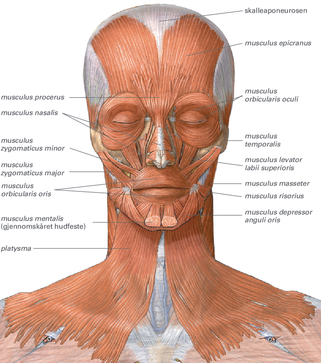
Musculus orbicularis oculi 1/5 Synonyms: Orbicularis oculi Orbicularis oculi is a flat, broad muscle that forms an ellipse around the circumference of the orbit. It is composed of orbital, palpebral and deep palpebral parts, each of which has its own specific set of attachments:

Orbicularis oculi muscle as face muscular system for eyelids outline diagram Muscular system
Orbicularis Oculi. The orbicularis oculi muscle surrounds the eye socket and extends into the eyelid. It has three distinct parts - palpebral, lacrimal, and orbital. Attachments - Originates from the medial orbital margin, the medial palpebral ligament, and the lacrimal bone. It inserts onto the skin around the margin of the orbit as well.
Orbicularis Oris Rehab My Patient
Orbicularis Oculi: the 2 blinking muscles We blink up to 20 times a minute and wink to convey complicity. This is thanks to the orbicularis oculi muscle, the subtle muscle keeping your eyes safe from bright lights, touch and foreign objects, even faster than you can think about it.
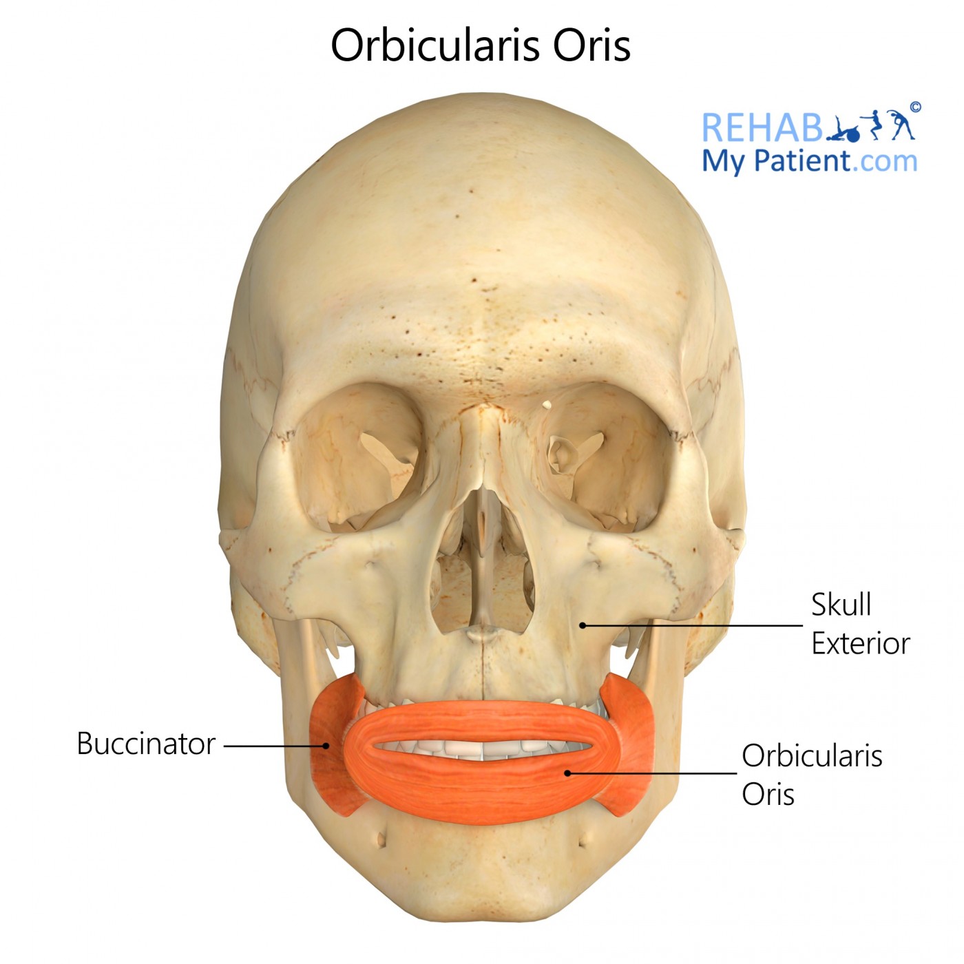
Orbicularis oris Origin, insertion, innervation, action Kenhub
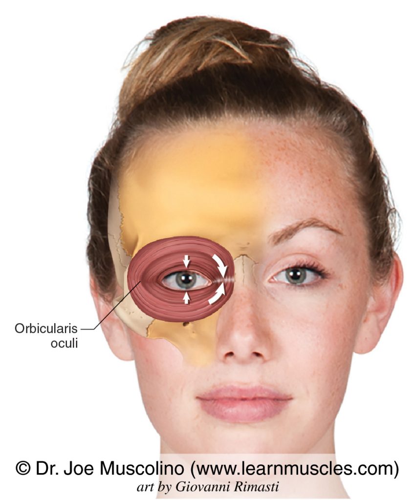
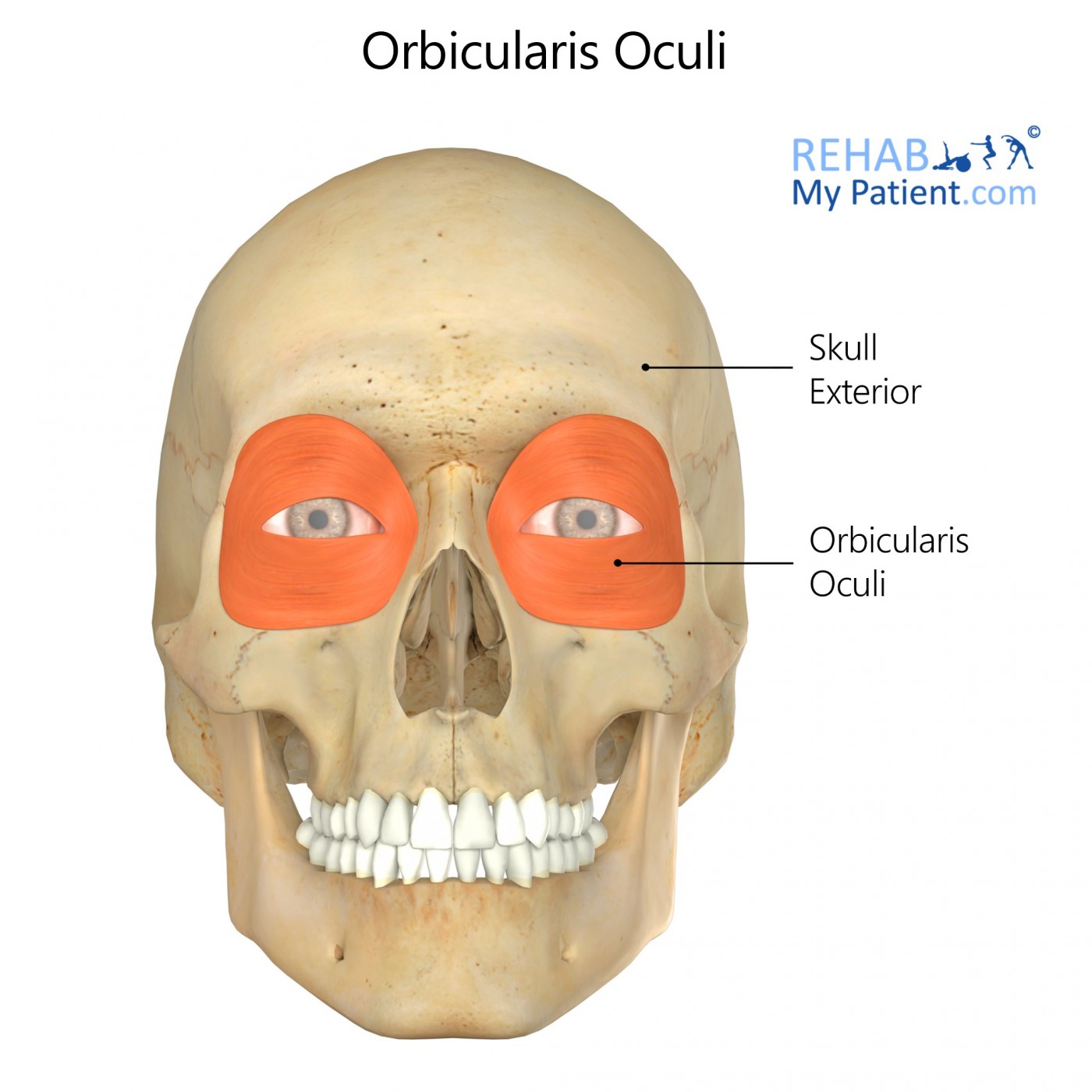
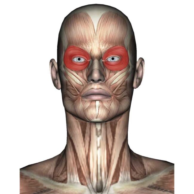
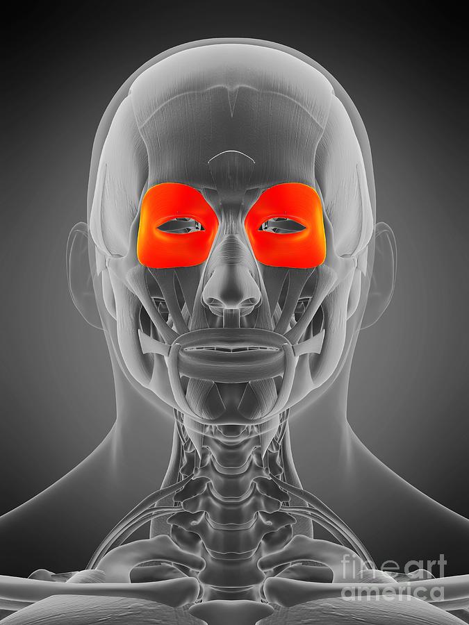
Orbicularis oculi. The orbicularis oculi (Latin: musculus orbicularis oculi) is a circular-shaped facial muscle located around the opening of the eye. This muscle is classified as the circumorbital and palpebral muscle. Among other functions, the orbicularis oculi muscle is involved in closing the eyelid. The orbicularis oculi is composed of.

The orbicularis oculi muscle Stock Image F002/1142 Science Photo Library
The orbicularis oculi receives innervation from the zygomatic and temporal branches of facial nerve (CN VII) and blood supply from branches of the maxillary, superficial temporal and facial arteries. The function of the orbicularis oculi depends on which part of the muscle contracts. Contraction of the orbital part pulls the skin of the.

Orbicularis oculi Location, Function and Pictures
The orbicularis oculi muscle is responsible for eyelid closure. It is a complex muscle, with a complicated spatial arrangement of myofibers. The muscle can be divided into three types: orbital, preseptal and pretarsal.

Orbicularis oculi muscle, illustration Stock Image F029/5057 Science Photo Library
Orbicularis oculi muscle - e-Anatomy - IMAIOS Human anatomy 2 Regions of human body Muscular system Muscles Fasciae Synovial bursae Tendon sheaths Cranial part of muscular system Muscles of head Extraocular muscles Superficial muscles of head Epicranius muscle Facial muscles Procerus muscle Nasalis muscle Depressor septi nasi
Orbicularis Oculi Definition, Function, Location, And Anatomy
The orbicularis oculi is a muscle in the face that closes the eyelids. It arises from the nasal part of the frontal bone, from the frontal process of the maxilla in front of the lacrimal groove, and from the anterior surface and borders of a short fibrous band, the medial palpebral ligament .
Orbicularis Oculi Rehab My Patient
The orbicularis oculi muscle, which is innervated by the facial nerve, is responsible for lid closure. It is subdivided into the pretarsal, preseptal, and orbital muscles. The orbicularis oculi is continuous with the superficial musculoaponeurotic system (SMAS) in the upper face as is the platysma in the lower face.
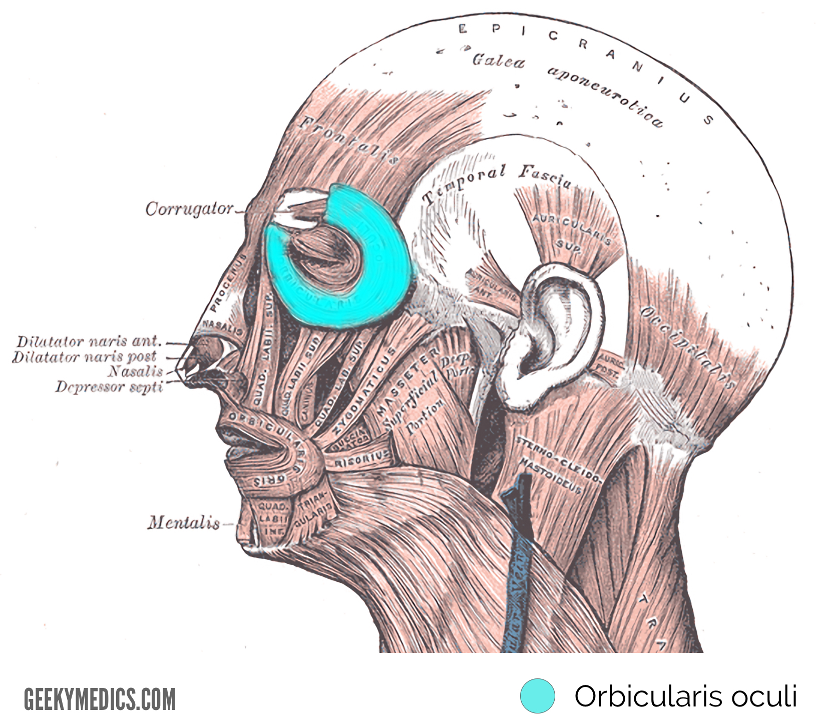
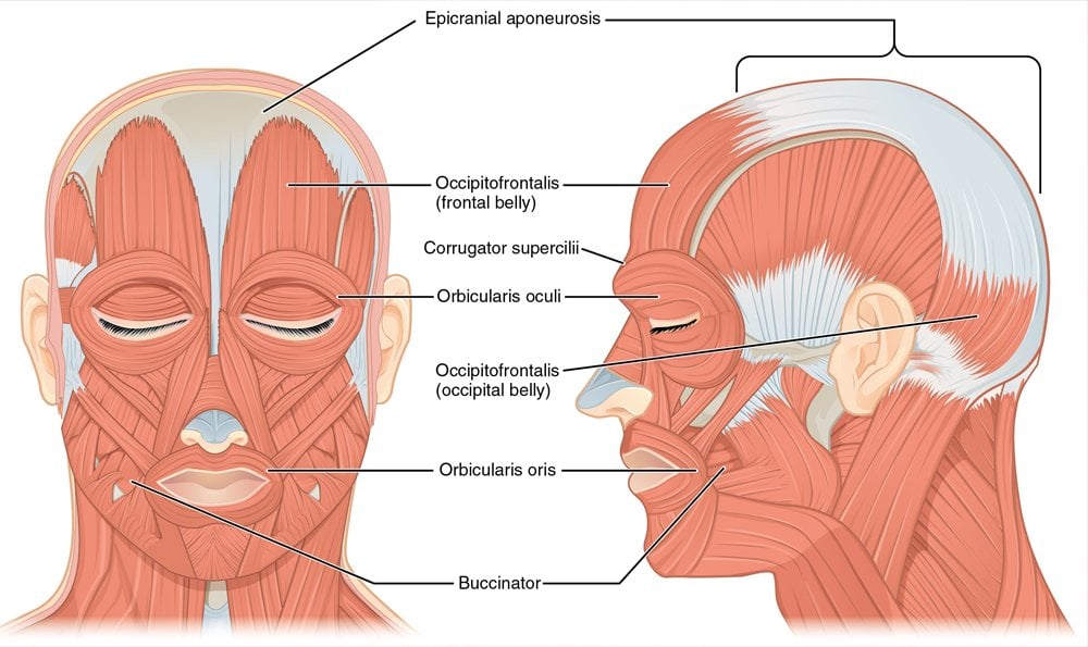
Muscles of Facial Expression Anatomy Geeky Medics
The orbicularis oculi muscle is innervated by the facial nerve, specifically the zygomatic and temporal branches. Surgical interventions may be necessary in cases of dysfunctional orbicularis oculi muscle, such as in the treatment of conditions like Bell's palsy or facial nerve injuries. These interventions aim to restore proper eyelid.
Orbicularis Oculi Anatomy Origin, Insertion, Action, Innervation The Wellness Digest
The orbicularis oculi muscle is a muscle surrounding each of the eyes and lies directly underneath the skin. The orbicularis oculi muscle is innervated by the cranial nerve VII, which means this.
Orbicularis Oculi Muscle Photograph by Sebastian Kaulitzki/science Photo Library Fine Art America
The orbicularis oculi muscle is a muscle of facial expression, a ring-like muscle functioning in a number of eyelid movements.. Gross anatomy. The orbicularis oculi muscle is subdivided into orbital, palpebral and lacrimal parts. Each has defined actions. The orbicularis oculi is secured to the medial and lateral palpebral ligament forming a ring in the eyelid tissue centered about the.
Orbicularis oculi Origin, insertion and action Kenhub
The orbicularis oculi is an orbital muscle of facial expression. It plays a key role in closing the eyelids and thus protecting the cornea from damage. Attachments - Originates from the medial orbital margin, the medial palpebral ligament, and the lacrimal bone.
musculus orbicularis oculi Store medisinske leksikon
Orbicularis oculi is one of the muscles of the eyelid. It is the primary sphincter muscle. It surrounds the orbit and extends out onto the temporal region and cheek. It consists of three parts which vary by location: orbital part. palpebral part. lacrimal part.

Orbicularis oculi (muscles of facial expression) Muscles of facial expression, Facial nerve
The orbicularis oculi muscle is a muscle located in the eyelids. It is a sphincter muscle arranged in concentric bands around the upper and lower eyelids. The main function of the orbicularis oculi muscle is to close the eyelids. This occurs when the muscle contracts. It also assists in the drainage of tears from the eyes.
